Foot & Ankle
What is the Normal Anatomy of the Foot and Ankle?
The foot and ankle form complex joints that are involved in movement and providing stability and balance to the body. The foot and ankle consist of 26 bones, 33 joints, and many muscles, tendons, and ligaments.
Bones of the Ankle
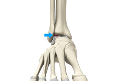
The ankle joint connects the leg with the foot and is composed of three bones: the tibia, fibula, and talus. The tibia or shinbone and fibula or calf bone are bones of the lower leg, which articulate with the talus or ankle bone, enabling up and down movement of the foot.
Three bony bumps present on the ends of the tibia and fibula form parts of the ankle joint:
- The medial malleolus, formed by the tibia, is found on the inside of the ankle.
- The posterior malleolus, also formed by the tibia, is found at the back of the ankle.
- The lateral malleolus, formed by the fibula, is found on the outer aspect of the ankle.
Bones of the Feet
The foot acts as a single functional unit, but can be divided into three parts: the hindfoot, midfoot and forefoot.
The hindfoot forms the ankle and heel, and is made up of the talus bone and calcaneus or heel bone. The heel bone is the largest bone in the foot.
The midfoot connects the hindfoot to the forefoot, and consists of one navicular bone, one cuboid bone, and three cuneiform bones. The navicular bone is found in front of the heel bone, and the cuneiform and cuboid bones are arranged in front of the navicular bone.
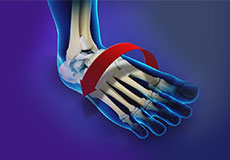
These bones are connected to five metatarsal bones of the forefoot that form the arch of the foot for shock absorption while walking or running. The forefoot is also made up of the toes or digits, formed by bones called phalanges - three in each toe, except the big toe, which has only two phalanges. The big toe has two additional tiny round sesamoid bones in the ball of the foot, which helps in upward and downward movements of the toe.
Ankle and Foot Joints
There are 33 joints in the ankle and foot. They include:
- Hinge joints in the ankle, which allow flexion (bending) and extension
- Gliding joints found in the hindfoot, which allow gliding movements
- Condyloid joints found in the forefoot and toes, which allow the flexion (bending) and extension, adduction, and abduction (sideward movement).
The joints of the foot and ankle provide stability and support the weight of your body, helping you to walk or run, and adapt to uneven grounds.
Soft Tissues of the Ankle and Foot
Our feet and ankle bones are held in place and supported by various soft tissues such as cartilage, ligaments, muscles, tendons, and bursae.
The joint surface of all the bones of the ankle and foot are lined by a thin, tough, flexible, and slippery surface called the articular cartilage, which acts as a shock absorber and cushion to reduce friction between the bones. The cartilage is lubricated by synovial fluid, which further enables smooth movement of the bones.
Ligaments are tough rope-like tissue that connect bones to other bones, and hold them in place, providing stability to the joints. The plantar fascia is the largest ligament in the foot, originating from the heel bone to the forefoot, it extends along the lower side of the foot and is involved in maintaining the arch of the foot. The plantar fascia ligament stretches and contracts to provide balance and strength to the foot. Lateral ligaments on the outside of the foot and medial ligaments on the inside of the foot provide stability and allow up and down movement of the foot.
The foot is made up of 20 muscles that help in movement. The main muscles include:
- Anterior tibial muscle, which allows up and down movement of the foot
- Posterior tibial muscle, which supports the arch
- Peroneal tibial muscle, which controls movement on the outside of the ankle
- Extensors, which enable the ankle to raise the toes just before stepping forward
- Flexors, which stabilize the toes against the floor
- Smaller muscles that help the toes to lift and curl
Tendons are soft tissues that connect muscles to bones. The largest and strongest tendon in the foot is the Achilles tendon, present at the back of the lower leg around the heel bone. Other tendons include peroneal and anterior and posterior tibialis.
Bursae are small fluid-filled sacs that decrease friction between tendons and bone or skin. They contain special cells called synovial cells that secrete a lubricating fluid.
Ankle Rheumatoid Arthritis
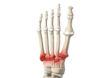
Rheumatoid arthritis is an auto-immune disease in which the body’s immune system (the body’s way of fighting infection) attacks its own healthy joints, tissues, and organs. It can cause pain, stiffness, swelling, and loss of function in the joints. Rheumatoid arthritis affects mostly joints of the hands and feet and tends to be symmetrical.
Bunionette
Bunionette also referred to as a tailor’s bunion is a bony lump that grows on the outside of the foot at the base of your little toe. The deformity got its name as q tailor’s bunion when tailors once sat with their legs crossed all day, with the outside edge of their feet rubbing on the ground.
Midfoot Arthritis
Midfoot arthritis is pain and inflammation of the midfoot. It occurs due to damage of cartilage or tissues around the joints. The damage may occur due to injury, aging or autoimmunity. The foot bones are the phalanges, the metaphalanges, and the tarsal bones.
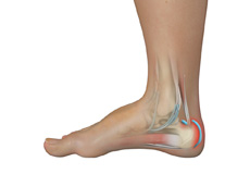
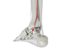
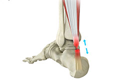
Achilles Tendon Rupture
The Achilles tendon is a strong fibrous cord present behind the ankle that connects the calf muscles to the heel bone. It is used when you walk, run and jump. The Achilles tendon ruptures most often in athletes participating in sports that involve running, pivoting and jumping.
Ankle Instability
The joints of the ankle are held in place and stabilized by strong bands of tissue called ligaments. Ankle instability is a chronic condition characterized by a recurrent slipping of the outer side of the ankle. It usually results from repeated ankle sprains, which are injuries to the ligaments. Ankle instability is generally noticed when you move your ankle joint but can also occur while standing.
Osteochondral Injuries of the Ankle
The ankle joint is formed by the articulation of the end of the tibia and fibula (shinbones) with the talus (heel bone). Osteochondral injuries, also called osteochondritis dissecans, are injuries to the talus bone. It is characterized by damage to the bone as well as the cartilage covering it.
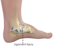
Ankle Ligament Injury
An ankle ligament injury, also known as an ankle sprain, can be caused by a sudden twisting movement of the foot during any athletic event or during daily activities. When stretched beyond its limit, the ligament may partially or completely tear. The injury can range from mild to severe, depending on the condition of the injured ligament and the number of ligaments involved.
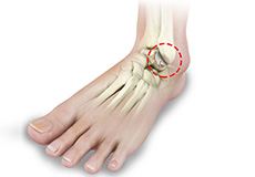
Foot and Ankle Arthritis
Arthritis is the inflammation of joints as a result of degeneration of the smooth cartilage that lines the ends of bones in a joint. This degeneration of the cartilages leads to painful rubbing of the bones, swelling, and stiffness in the joints, resulting in restricted movements. Arthritis in the foot and ankle can occur due to fractures, dislocation, inflammatory disease, or congenital deformity.
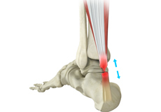
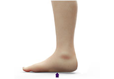
Achilles Tendon Bursitis
Achilles tendon bursitis or retrocalcaneal bursitis is a condition that commonly occurs in athletes. It is a painful condition caused by the swelling of the bursa, a fluid-filled sac that is located at the back of the heel under the Achilles tendon. This retrocalcaneal bursa contains a lubricating fluid that acts as a cushion to reduce friction between muscle and bones.
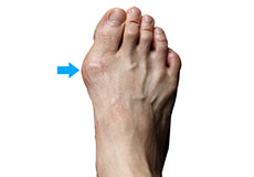
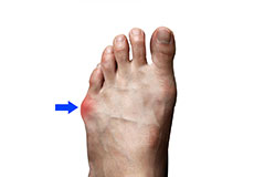
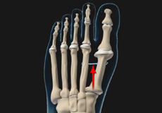
Bunion
A bunion is a bony protuberance that appears on the outer surface of the big toe when it angles toward the adjacent toe. It is an extra bone and a fluid-filled sac that grows at the base of the big toe.
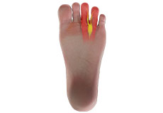
Morton's Neuroma
Morton’s neuroma refers to a nerve injury that occurs between the toes, usually the third and fourth toes. This causes pain and thickening of the nerve tissue.
Flatfoot
Flatfoot, also known as “fallen arches” or Pes planus, is a deformity in children’s feet where the arch that runs along the sole of the foot collapses to the ground or is not formed at all. Flatfoot is normal in the first few years of life as the arch of the foot usually develops between the age of 3 and 5 years.
Posterior Tibial Tendon Dysfunction
The posterior tibial tendon passes through the ankle to attach the calf muscle with the bones of the midfoot. It provides stability to the arch and supports the foot while walking. Inflammation or a tear of this tendon as a result of injury may cause dysfunction, leading to pain and the development of a flatfoot.
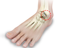
Osteochondral Lesions of the Ankle
The tibia and the fibula bones of the lower leg join with the talus bone to form the ankle joint. The talus bone is an important bone located between the tibia and fibula and the heel bone (calcaneus). OCL or OCD is the damage to the cartilage and the talus bone of the ankle joint. Usually, the inner or the medial portion of the ankle is affected.
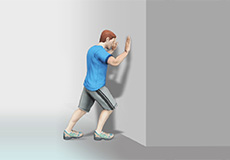
Foot Rehabilitation Following Surgery
A foot injury or foot surgery may leave you immobile for a period of time. To return to your regular activities and more strenuous recreational activities, it is necessary for you to follow a well-planned activity and exercise program.
Foot Reconstruction
Foot reconstruction is a surgery performed to correct the structures of the foot and restore the natural functionality of the foot that has been lost due to injury or illness. Ideally, any foot surgery for reconstruction is done to improve the appearance and function of the foot so that you can maintain your quality of life.
Ankle Joint Replacement
The ankle joint connects the leg with the foot and provides free movement to the foot. It is formed by connecting the bones of the lower leg, tibia, and fibula, with the talus, or ankle bone.
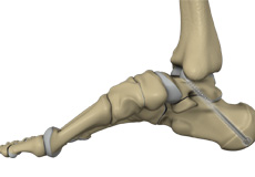
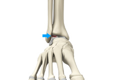
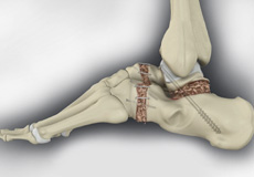
Subtalar Arthrodesis
Subtalar arthrodesis is the surgical fusion of bones that form the subtalar joint. The goal of subtalar arthrodesis is to relieve pain in the affected joint. This is achieved by surgically eliminating the joint.
Ankle Arthroscopy
Ankle arthroscopy is a minimally invasive surgical procedure in which an arthroscope, a small, soft, flexible tube with a light and video camera at the end, is inserted into the ankle joint to evaluate and treat a variety of conditions. The camera projects an image of the inside of the joint onto a large monitor, allowing your surgeon to look for any damage, assess the type of injury and repair the problem.
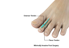
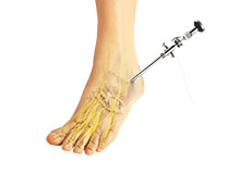
Minimally Invasive Foot Surgery
Minimally invasive foot surgery (MIFS) uses the latest advanced technology to treat foot and ankle pain caused by a variety of conditions. Special surgical instruments, devices, and advanced imaging techniques are used to visualize and perform the surgery through small incisions.
Flatfoot Reconstruction
Foot reconstruction is a surgery performed to correct the structures of the foot and restore the natural functionality of the foot that has been lost due to injury or illness. Flatfoot or pes planus is a condition in which the foot does not have a normal arch when standing.
Achilles Tendon Repair
The Achilles tendon is often injured during sports activities, resulting in an inflammatory condition called tendonitis, which is characterized by swelling and pain. In some cases, severe injury results in a tear or rupture of the Achilles tendon, requiring immediate medical attention.
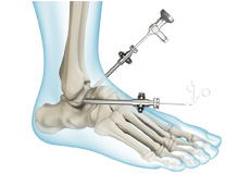
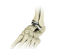
Ankle Arthrodesis
Ankle arthrodesis is the surgical fusion of bones that form the ankle joint. The ankle joint is formed by the tibia, talus, and the fibula bones.

Ankle Ligament Reconstruction
Ankle ligament reconstruction may be performed arthroscopically under general anesthesia. Your surgeon will make small incisions in your ankle. A tiny camera and a few special instruments are inserted through the incisions to repair and strengthen the ligaments. Stretched or torn ligaments will be shortened and stitched as needed.
Ankle Instability Surgery
Ankle instability is a chronic condition characterized by the recurrent slipping of the outer side of the ankle. Instability is generally noticed during movement of the ankle joint, but can also occur while standing.
Bunionectomy
A bunionectomy is a surgical procedure to remove a bunion. A bunion, also called a hallux valgus, is an enlargement of bone or soft tissues around the joint at the base of the big toe that results in the formation of a bump.
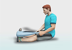
Foot and Ankle Rehabilitation
The foot is composed of different structures including bones, ligaments, tendons, and muscles. As the feet bear the weight of our body, they are more prone to injury and pain. A foot injury or foot surgery may leave you immobile for a period of time.