Hip
Hip Joint
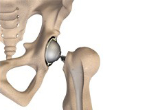
The hip joint is the largest weight-bearing joint in the human body. It is also referred to as a ball and socket joint and is surrounded by muscles, ligaments, and tendons. The thigh bone or femur and the pelvis join to form the hip joint.
Any injury or disease of the hip will adversely affect the joint's range of motion and ability to bear weight.
The hip joint is made up of the following:
- Bones and joints
- Ligaments of the joint capsule
- Muscles and tendons
- Nerves and blood vessels that supply the bones and muscles of the hip
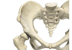
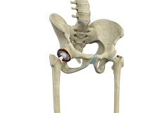
Bones and Joints
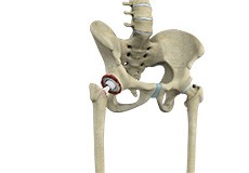
The hip joint is the junction where the hip joins the leg to the trunk of the body. It is comprised of two bones: the thigh bone or femur and the pelvis which is made up of three bones called ilium, ischium, and pubis. The ball of the hip joint is made by the femoral head while the socket is formed by the acetabulum. The Acetabulum is a deep, circular socket formed on the outer edge of the pelvis by the union of three bones: ilium, ischium, and pubis. The lower part of the ilium is attached by the pubis while the ischium is considerably behind the pubis. The stability of the hip is provided by the joint capsule or acetabulum and the muscles and ligaments which surround and support the hip joint.
The head of the femur rotates and glides within the acetabulum. A fibrocartilagenous lining called the labrum is attached to the acetabulum and further increases the depth of the socket.
The femur or thigh bone is one of the longest bones in the human body. The upper part of the thigh bone consists of the femoral head, femoral neck, and greater and lesser trochanters. The head of the femur joins the pelvis (acetabulum) to form the hip joint. Next, to the femoral neck, there are two protrusions known as greater and lesser trochanters which serve as sites of muscle attachment.
Articular cartilage is the thin, tough, flexible, and slippery surface lubricated by synovial fluid that covers the weight-bearing bones of the body. It enables smooth movements of the bones and reduces friction.
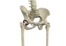
Ligaments
Ligaments are fibrous structures that connect bones to other bones. The hip joint is encircled with ligaments to provide stability to the hip by forming a dense and fibrous structure around the joint capsule. The ligaments adjoining the hip joint include:
Iliofemoral ligament: This is a Y-shaped ligament that connects the pelvis to the femoral head at the front of the joint. It helps in limiting the over-extension of the hip.
Pubofemoral ligament: This is a triangular shaped ligament that extends between the upper portion of the pubis and the iliofemoral ligament. It attaches the pubis to the femoral head.
Ischiofemoral ligament: This is a group of strong fibers that arise from the ischium behind the acetabulum and merge with the fibers of the joint capsule.
Ligamentum teres: This is a small ligament that extends from the tip of the femoral head to the acetabulum. Although it has no role in hip movement, it does have a small artery within that supplies blood to a part of the femoral head.
Acetabular labrum: The labrum is a fibrous cartilage ring which lines the acetabular socket. It deepens the cavity, increasing the stability and strength of the hip joint.
Muscles and Tendons
A long tendon called the iliotibial band runs along the femur from the hip to the knee and serves as an attachment site for several hip muscles including the following:
Gluteals: These are the muscles that form the buttocks. There are three muscles (gluteus minimus, gluteus maximus, and gluteus medius) that attach to the back of the pelvis and insert into the greater trochanter of the femur.
Adductors: These muscles are located in the thigh which helps in adduction, the action of pulling the leg back towards the midline.
Iliopsoas: This muscle is located in front of the hip joint and provides flexion. It is a deep muscle that originates from the lower back and pelvis and extends up to the inside surface of the upper part of the femur.
Rectus femoris: This is the largest band of muscles located in front of the thigh. They also are hip flexors.
Hamstring muscles: These begin at the bottom of the pelvis and run down the back of the thigh. Because they cross the back of the hip joint, they help in extension of the hip by pulling it backward.
Nerves and Arteries
Nerves of the hip transfer signals from the brain to the muscles to aid in hip movement. They also carry the sensory signals such as touch, pain, and temperature back to the brain.
The main nerves in the hip region include the femoral nerve in the front of the femur and the sciatic nerve at the back. The hip is also supplied by a smaller nerve known as the obturator nerve.
In addition to these nerves, there are blood vessels that supply blood to the lower limbs. The femoral artery, one of the largest arteries in the body, arises deep in the pelvis and can be felt in front of the upper thigh.
Hip Movements
All of the anatomical parts of the hip work together to enable various hip movements. Hip movements include flexion, extension, abduction, adduction, circumduction, and hip rotation.
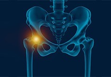
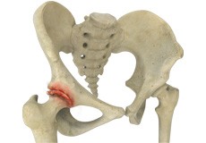
Hip Pain
Hip pain, one of the common complaints, may not always be felt precisely over the hip joint rather in and around the hip joint. The cause for pain is multifactorial and the exact position of your hip pain suggests the probable cause or underlying condition causing it. Pain felt inside the hip joint or your groin area is more likely to be because of the problems within the hip joint.
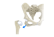
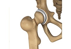
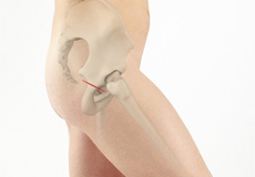
Hip Dislocation
The hip joint is a “ball and socket” joint. The “ball” is the head of the femur or thighbone, and the “socket” is the cup-shaped acetabulum. The joint is surrounded by muscles, ligaments, and tendons that support and hold the bones of the joint in place. Hip dislocation occurs when the head of the femur moves out of the socket.
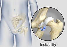
Hip Instability
Injury or damage to these structures can lead to a condition called hip instability when the joint becomes unstable. Hip instability can be traumatic or atraumatic. Traumatic instability can be caused by injuries from sports or motor vehicle accidents.
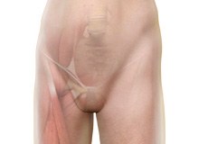
Hip Groin Disorders
Hip and groin disorders are more common in athletes. They are caused by rapid acceleration and deceleration motion. The rehabilitation time for hip and groin injuries is longer than most other injuries, therefore, early and accurate diagnosis is essential.
Developmental Dysplasia
Developmental dysplasia of the hip (DDH) or hip dysplasia is a condition that is seen in infants and young children because of developmental problems in the hip joint. The femur (thighbone) partially or completely slips out of the hip socket leading to dislocation at the hip joint. It is most common in the first-born baby with a family history of the disorder.
Osteoarthritis of the Hip
Osteoarthritis, also called degenerative joint disease, is the most common form of arthritis. It occurs most often in the elderly. This disease affects the tissue covering the ends of bones in a joint called cartilage. In osteoarthritis, the cartilage becomes damaged and worn out, causing pain, swelling, stiffness and restricted movement in the affected joint.
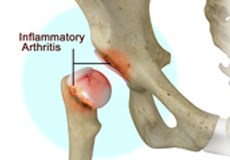
Inflammatory Arthritis of the Hip
The inflammation of the joints is referred to as arthritis. Inflammation arises when the smooth lining called cartilage at the ends of bones wears away. In some cases, the inflammation is caused when the lining of the joint becomes inflamed as part of an underlying systemic disease. These conditions are referred to as inflammatory arthritis.
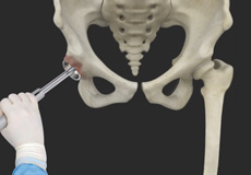
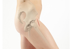
Mini-Posterior Hip Replacement
Mini-posterior hip replacement is a surgical procedure used to replace your damaged hip with synthetic parts inserted through a small incision made at the back of the hip.
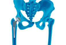
Robotic Total Hip Replacement
Total hip replacement is a surgical technique where the severely damaged cartilage and bone of the hip joint are removed and substituted with an artificial prosthesis which is bio-compatible and functions like a normal hip. It is one of the most common joint replacement procedures, subsequent to knee replacements.
Posterior Hip Replacement
Posterior hip replacement is a minimally invasive hip surgery performed to replace the hip joint. It is also referred to as muscle sparing surgery because no muscles are cut to access the hip joint, enabling a quicker return to normal activity.
Outpatient Anterior Approach Hip Replacement
With improved technology and advances in anesthesia and pain control, hip replacement surgery has evolved and is now being offered in an outpatient setting.
Correction of a Failed Hip Replacement
Reoperation of a total hip replacement to resolve a painful hip condition arising out of a damaged or worn out prosthesis (artificial hip joint) is known as correction of a failed hip replacement. During this corrective surgery, a partial or complete exchange of the prostheses that were implanted during the original surgery is done.
Correction of a Painful Hip Replacement
Reoperation of a total hip replacement to resolve a painful hip condition arising out of a damaged or worn out prosthesis (artificial hip joint) is known as correction of a painful hip replacement. During this corrective surgery, a partial or complete exchange of the prostheses that were implanted during the original surgery is done.
Correction of a Loose Hip Replacement
Reoperation of a total hip replacement to resolve a painful hip condition and loss of motion due to a loosened prosthesis (artificial hip joint) is known as correction of a loose hip replacement. This loosening occurs due to wear and tear of the implant surfaces and subsequent weakening of the surrounding bone. During this corrective surgery, a partial or complete exchange of the prostheses that were implanted during the original surgery is done.
Outpatient Hip Replacement
Hip replacement surgery is one of the most common orthopedic surgeries performed. It involves the replacement of the damaged hip bone (ball shaped upper end of the femur) with a ceramic ball attached to a metal stem that is fixed into the femur and placing a new cup with a special liner in the pelvis.
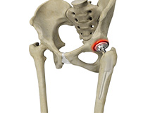
Total Hip Replacement
Total hip replacement is a surgical procedure in which the damaged cartilage and bone is removed from the hip joint and replaced with artificial components.
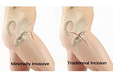
Minimally Invasive Total Hip Replacement
Surgery may be recommended in patients with severe cartilage damage and if conservative treatment options such as anti-inflammatory medications and physical therapy do not relieve the symptoms.
Anterior approach hip replacement
Anterior approach hip replacement surgery is performed under general anesthesia or regional anesthesia. You will lie down on your back, on a special operating table that enables your surgeon to perform the surgery from the front of the hip. Your surgeon may use fluoroscopic imaging during the surgery to ensure the accuracy of component positioning and to minimize leg length inequality.
Revision Hip Replacement
During total hip replacement, the damaged cartilage and bone are removed from the hip joint and replaced with artificial components. At times, hip replacement implants can wear out for various reasons and may need to be replaced with the help of a surgical procedure known as revision hip replacement surgery.
Computer-assisted Hip Replacement
Computer-assisted hip replacement is an image-guided, minimally invasive surgical procedure to replace your diseased or damaged hip with an artificial device using the assistance of computer software. The system creates and displays images and provides information that aids your surgeon at various stages of the procedure to improve accuracy and results.
Computer-Navigated Total Hip Replacement
For a successful total hip replacement, accurate positioning of the implants is crucial to accomplish a good clinical outcome. Computer-navigated total hip replacement is an advanced technology developed to provide more accurate positioning of an implant. Hip replacement through computer navigation provides information and guidance to the surgeon for precise positioning of implants.
Direct Superior Hip Replacement
Direct superior hip replacement is a minimally invasive surgery that provides your surgeon access to the hip joint with minimum displacement or damage to the surrounding muscles, tendons, and ligaments.
Hip Reconstruction
Hip reconstruction is a surgery to repair or replace a damaged hip joint that causes pain and limits your movement.
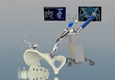
Robotic Assisted Hip Surgery
Robotic assisted hip surgery is a minimally invasive surgical procedure that involves the use of a specialized robotic system to remove the damaged parts of a hip joint and replace them with an artificial prosthesis or implant.
Same Day Hip Replacement Surgery
Hip replacement surgery is one of the most common orthopedic surgeries performed. It involves the replacement of the damaged hip bone (ball-shaped upper end of the femur) with a ceramic ball attached to a metal stem that is fixed into the femur and placing a new cup with a special liner in the pelvis.

Muscle Sparing Hip Replacement
Muscle sparing anterior hip replacement is a minimally invasive hip surgery to replace the hip joint without cutting through any muscles or tendons as compared to traditional hip replacement that involves cutting major muscles to access the hip joint.

Activities After Hip Replacement
Hip replacement is a surgery performed to replace parts of a diseased hip joint with a prosthesis. The goal of hip replacement is to eliminate pain and enable you to return to your normal activities. You can help in the recovery and improve the outcomes of the procedure by following certain precautions and changing the way you carry out your daily activities.